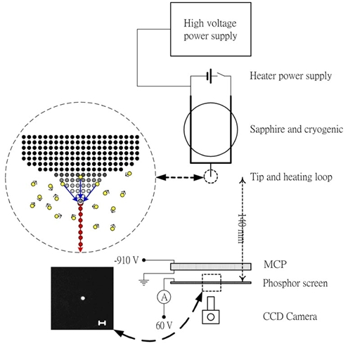
A. We have developed a new, simple and reproducible method for preparing noble metal-covered W(111) single-atom tips. By electroplating a noble metal (Pd, Pt, Rh, Ir, Au) thin film on single-crystal W (111) tips followed by thermal annealing in vacuum, single-atom tips can be obtained. These tips are both thermally and chemically stable, and the same tip structure can also be regenerated even if damaged accidentally. The electron field emission characteristics were also investigated. The brightness is estimated to be at least one order of magnitude higher than that of normal tungsten emitters or Schottky emittere, which are the state-of-the-art electron sources in current electron microscopes. The small opening angle, smallest emitting area, high brightness, high wave coherence, and high stability of the electron beams field emitted from these single-atom tips make their future commercial application to electron microscopy, electron spectroscopy, and electron holography, and other electron-beam based advanced instrumentations become possible. (NANO Letters 4(12), 2379 (2004))
B. We have demonstrated that an Iridium covered W(111) single-atom tip can be a good field emitter for helium, hydrogen, oxygen, or argon gas ion source. The brightness of these gas field ion sources (GFISs) is at least one order of magnitude better than that of liquid-metal ion sources used in current commercial focused ion beam (FIB) systems. The measured ion current stability and the lifetime of these GFISs are also good enough for applications in FIB systems. (Appl. Phys. Lett. 92, (2008) 063106)
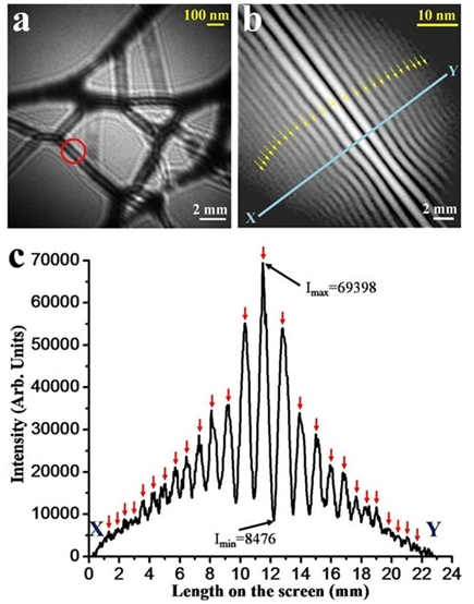
C. We have demonstrated that the electron beam from a noble metal-covered W(111) single-atom tip is fully spatially coherent. This property may lead to the use of phase coherence of electron beam in the future. Information about the electric and magnetic fields in a material down to atomic scale might be retrieved with the use of the fully coherent electron sources. We have also used the coherent electron beam emitted from our single-atom tips to image single-walled carbon nanotubes using a home-made low-energy electron point projection microscope (PPM). Through cooperation with a theoretical group, the mechanism for the image contrast and the interference fringes in PPM are clarified for the first time. (Nanotechnology 20, 115401 (2009))