Spherical microwell arrays for studying single cells and microtissues in 3D confinement
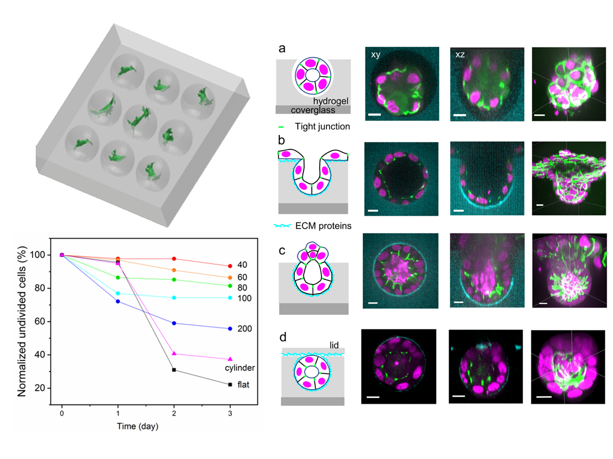
Microwell arrays have emerged as three-dimensional substrates for cell culture due to their simplicity of fabrication and promise for high-throughput applications such as 3D cell-based assays for drug screening. To date, most microwells have had cylindrical geometries. Motivated by our previous findings that cells display 3D physiological characteristics when grown in the spherical micropores of monodisperse foam scaffolds [1, 2], here we engineered novel microwells shaped as spherical caps with obtuse polar angles, yielding narrow apertures. When used as bare substrates, these microwells were suitable for culturing cell spheroids; the narrow apertures sterically hindered unattached cultured cells from rolling out of microwells under agitation. When only the walls of the microwell were conjugated with extracellular matrix proteins, cells remained confined in the microwells. Epithelial cells proliferated and burst out of the aperture, and cell polarity was oriented based on the distribution of extracellular matrix proteins in the microwells. Surprisingly, single fibroblast cells in spherical wells of various diameters (40-100 µm) underwent cell-cycle arrest, while cells in circular cylindrical microwells continued to proliferate. Spatial confinement was not sufficient to cause cell-cycle arrest; however, confinement in a constant negative-curvature microenvironment led to cell-cycle arrest. Overall, these investigations demonstrate that this spherical microwell substrate constitutes a novel basic research tool for elucidating how cells respond to dimensionality and microenvironment with radii of curvature at the cellular length scale.