3D chemical imaging of the brain using quantitative IR spectro-microscopy
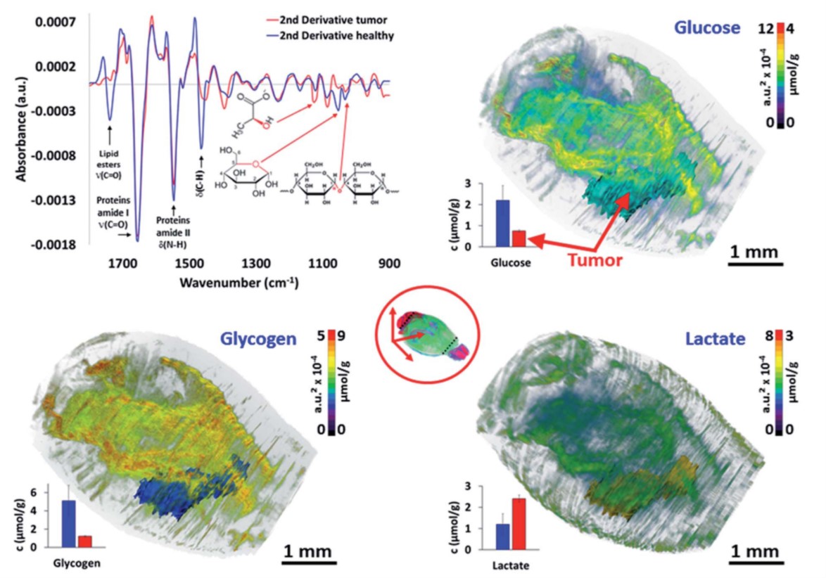
Three-dimensional (3D) histology is the next frontier for modern anatomo-pathology. Characterizing abnormal parameters in a tissue is essential to understand the rationale of pathology development. However, there is no analytical technique, in vivo or histological, able to discover such abnormal features and provide their 3D distribution at microscopic resolution. Here, we introduce a unique high-throughput infrared (IR) microscopy method, which combines automated image corrections and subsequent spectral data analysis for 3D-IR image reconstruction. We performed the spectral analysis of a complete organ for a small animal model, a mouse brain with an implanted glioma tumor. The 3D-IR image is reconstructed from 370 consecutive tissue sections and corrected with the X-ray tomogram of the organ for an accurate quantitative analysis of chemical contents. A 3D matrix of 89.106 IR spectra is generated, allowing to separate the tumor mass from healthy brain tissues based on various anatomical, chemical, and metabolic parameters. We demonstrate that quantitative metabolic parameters can be extracted from IR spectra for characterization of brain vs. tumor metabolism (assessing the Warburg effect in tumor). Our method can be further exploited by searching for whole spectral profile discriminating tumor vs. healthy tissue in a non-supervised manner, which we call ‘spectromics’. The complete results have been published in the Chemical Science.
WebSite: http://pubs.rsc.org/en/content/articlelanding/2017/sc/c7sc03306k#!divAbstract
WebSite: http://pubs.rsc.org/en/content/articlelanding/2017/sc/c7sc03306k#!divAbstract